Saturday, September 17, 2016

3.17 Understand the meaning of the terms: dominant, recessive, homozygous, heterozygous, phenotype, genotype and codominance

Homozygous: two same alleles of a gene on each chromosome on a homologous pair
Heterozygous: one of each allele of a gene on each chromosome on a homologous pair
Rescessive: an allele only expressed if the dominant allele is absent or if [the recessive gene] is homozygous.
Dominant: an allele that always express itself whether it is heterozygous or homozygous


Homologous pair: a chromosome containing a maternal and paternal chromatid joined together. (these have the same gene but may have different alleles)
Chromatid: each of the two threadlike strands that chromosomes divide into during mitosis.


3.16 Understand that genes exist in alternative forms called alleles which give rise to differences in inherited characteristics

Genes exist in alternative forms called alleles. These control the differences in inherited characteristics. 

Extra information:
Alleles can be dominant or recessive, homozygous or heterozygous. They are written as big or small letters. Eg: Aa or AA or aa. This is known as a genotype. The physical characteristic it codes for is called a phenotype.

3.15 Describe a DNA molecule as two strands coiled to form a double helix, the strands being linked by a series of paired bases: adenine (A) with thymine (T), and cytosine (C) with guanine (G)

Above, you can see an image of the structure of DNA. It is made up of two coiled strands, known as a sugar phosphate backbone. As the name suggests, it is made up of a sugar and a phosphate (alternating). The base pairs are the 'coding'. It is believed that every living thing has the same base pairs:

  • Adenine (A)
  • Thymine (T)
  • Guanine (G)
  • Cytosine (C)
Adenine always pairs up with thymine, guanine always pairs up with cytosine. The order in which these are paired in is what makes up the 'coding' of an organism.

(Virtual flashcards on this topic can be found here, or click 'inheritance flashcards' on the right hand side menu)

Friday, September 16, 2016

3.14 Understand that a gene is a section of a molecule of DNA and that a gene codes for a specific protein

A gene is a length of DNA. This codes for a specific protein. These proteins join together to make a chain. 

Structure of DNA

3.13 Understand that the nucleus of a cell contains chromosomes on which genes are located


The nucleus of any cell will contain chromosomes in which genes are located. 
If you unwrapped a chromosome, you would see it is essentially just strands of DNA coiled around a protein. DNA stands for deoxyribonucleic acid (you don't need to know that).

Wednesday, June 1, 2016

4.7 Explain why only about 10% of energy is transferred from one trophic level to the next.


  • Some energy is used at the current trophic level in life processes
  • Some food the organism takes up is undigested and passed as faeces
  • Organism might not eat everything (i.e. a rabbit won't eat a cabbage's roots, so won't gain 100% of its energy even if no energy was used by the cabbage)

4.6 Understand the transfer of substances and of energy along a food chain

When one organism eats another, the substances and energy from it get transferred to its consumer. I.e. if you eat a kilo of spinach, the energy and nutrients from that are transferred to you (don't try that please)

2.90 understand the sources, roles and effects of the following hormones: ADH, adrenaline, insulin, testosterone, progesterone and oestrogen

Basically everything you need to know is above, sorry if my handwriting is unreadable and messy ↑

Tuesday, May 31, 2016

2.84 Understand that the central nervous system consists of the brain and spinal cord and is linked to sense organs by nerves

The central nervous system consists of:

  • brain
  • spinal cord
and is linked to sense organs (i.e. eyes, tongue, touch receptors) by nerves

2.83 Describe how responses can be controlled by nervous or by hormonal communication and understand the differences between the two systems

Endocrine (hormonal) system

  • Long lasting response
  • Widespread effect
  • Hormones produced all over the body, carried by plasma
  • Pituitary gland and hypothalamus are key players
Nervous
  • Short lived but very quick
  • Localised; specific to an area
  • Often involves the contraction of a muscle

2.56 Describe experiments to investigate the role of environmental factors in determining the rate of transpiration from a leafy shoot


  • Weigh the plant at the start
  • Put it in a test tube with only its roots / end in the water
  • Put a layer of oil over water
  • Leave for x amount of time
  • Weigh plant again
  • Record results
  • Repeat, changing one variable (i.e temperature)

2.55 Explain how the rate of transpiration is affected by changes in humidity, wind speed, temperature and light intensity


Humidity
Decreases amount of transpiration because the concentration of H₂O in the air will equal that of the plant, so it can't move from high to low concentration as per usual 

Wind
Increases transpiration because water is blown away, which means there is a greater difference in H₂O concentration

Temperature
Increases transpiration: the heat makes the water evaporate more quickly.

Light intensity
Stronger light means more transpiration because leaves absorb more heat from the light, so water evaporates quicker

2.54 understand that transpiration is the evaporation of water from the surface of a plant

Transpiration is when water evaporates from the surface of a plant; typically below the leaf because of the stomata being opened by guard cells

2.53 Explain how water is absorbed by root hair cells


  • Water is absorbed by osmosis
  • (Minerals absorbed by active transport)
  • Hair cells are long in shape, giving it a large surface area (also, they branch)
  • Epidermal cells increase surface area further
  • Makes osmosis faster and more efficient
  • Substances are transported by the xylem

4.5 Understand the concepts of food chains, food webs, pyramids of number, pyramids of biomass and pyramids of energy transfer

Food chain
Shows energy transfer between producers all the way to tertiary consumers. Only 1 line
eg: grass > zebra > lion

Food web
Shows links between various food chains within the same habitat or environment
eg:

Pyramid of numbers
Shows number of organisms in each trophic level and isn't always a nice pyramid shape
eg:

Pyramid of biomass
Shows mass of organism in each trophic level, and is almost always a pyramid shape
eg:

Energy transfer pyramid / chain
Shows energy transfer between organisms, typically in a food chain
eg


4.4 Explain the names given to different trophic levels to include producers, primary, secondary and tertiary consumers and decomposers

Producers
Produce their own 'food' (glucose) from the sun's light energy

Primary consumer
Eats (consumes) producer and gains its energy; often a herbivore

Secondary consumer
Eats (consumes) primary consumer and gains its energy; often an omnivore

Tertiary consumer
Eats secondary consumer and gains its energy; top of the food chain; often a carnivore

When these die, they're broken down by fungi and bacteria. Nitrogen and carbon compounds are returned to the earth (see cycles)


4.3 Explain how quadrats can be used to sample the distribution of organisms in their habitats.


  • Quadrats placed in different areas at random
  • One is chosen
  • Population counted (must be either plants or VERY slow moving animals i.e. slug)
  • Repeated in different areas
  • Population counted
  • You can compare results and see where population is more dense (i.e. there might be more flowers near a river bed)

4.2 Explain how quadrats can be used to estimate the population size of an organism in two different areas

Sample area is taken by quadrat, can be repeated many many times to make it more accurate, then multiplied to 'cover' desired area of land. 

4.1 Understand the terms population, community, habitat and ecosystem


  • Population: number of a certain type or types or organism(s) living in a particular area at a particular time
  • Community: Interacting group of various species in a common location: typically relying on one another
  • Habitat: physical location of an organism, including what is available to it
  • Environment: the natural biological factors that affect a species or particular organism (i.e. humans)

2.67 Understand the origin of carbon dioxide and oxygen as waste products of metabolism and their loss from the stomata of a leaf

Both CO₂ and O₂ are waste products of metabolic reactions:

  • CO₂ + H₂O > C₆H₁₂O₆ + O₂ (photosynthesis)
  • C₆H₁₂O₆ + O₂>  CO₂ + H₂O (respiration)
which are carried out by plants. 

2.52 Describe the role of xylem in transporting water and mineral salts


  • Hollow, dead cells
  • Transports water and mineral salts
  • Side by side with phloem
  • EG Nitrates, phosphates, etc.

2.51 Describe the role of phloem in transporting sucrose and amino acids between the leaves and other parts of the plant


  • Phloem has living cells in form of a column
  • Has companion cells and a cytoplasm + cellulose cell wall
  • Companion cells very metabolically active
  • Carries dissolved substances such as sucrose and amino acids within the plant

2.50 Understand the need for a transport system in multicellular organisms

Multicellular organisms have a small surface area to volume ratio so diffusion is insufficient and would be too slow anyway. Transport systems speed up the process of getting the molecules required (i.e circulatory)

2.49 Understand why simple, unicellular organisms can rely on diffusion for movement of substances in and out of the cell

They have a very high surface area to volume ratio, so things diffuse in and out very quickly. Also, they are so small they don't use up that much in terms of energy, food, etc. so diffusion is sufficient.

2.43 Describe experiments to investigate the effect of light on net gas exchange from a leaf, using hydrogen-carbonate indicator

As light increases, so does photosynthesis. A hydrogen carbonate indicator, like limewater, is an indicator for CO₂. 


  • Normal: orange
  • Increase: yellow
  • Decrease: purple

  • Fill a test tube 1/4 full with HCl. 
  • Attach leaf to bung and leave it in there
  • Vary light intensity every x minutes
  • (Change distance y from leaf)
  • Record results
  • done

Monday, May 30, 2016

2.22 Describe experiments to investigate photosynthesis, showing the evolution of oxygen from a water plant, the production of starch and the requirements of light, carbon dioxide and chlorophyll

Pond weed experiment
  • Pond weed in large beaker with water
  • Count bubbles released in 1 minute
  • Record results and repeat 3x
  • Repeat, changing either temperature, concentration of CO2, light intensity and / or plant species
Image of apparatus

2.42 Describe the role of stomata in gas exchange

Stomata is just the plural form of 'stoma', which are the tiny pores on the underside of the leaf. Guard cells control whether they open or close, and therefore also control the exchanging of carbon dioxide and oxygen. I'm pretty sure I already covered how they work in another post, but I'll just write it again ... (bear with me)

If there are a lot of potassium ions present in the fluid in the guard cells, the cells will absorb a lot of water via osmosis, and will become more turgid. This opens the stoma, allowing diffusion to take place. If there aren't a lot of potassium ions present, the cells will lose water, become flaccid and close the stoma. The opening and closing of the stoma also depends on whether it is day or night. In the light, the guard cells absorb water, in the dark they lose it.

2.41 Explain how the structure of the leaf is adapted for gas exchange


  • The cells in the spongy mesophyll (lower layer) are loosely packed, which increases the surface area of the leaf, allowing for more carbon dioxide to be absorbed
  • Guard cells (stomata) on the lower part of the leaf controls the diffusion of gases in to the leaf.
  • Leaves are thin, making it easier and quicker for gas to diffuse quickly through it.
Let me explain how the stomata work: 
- During the day, the guard cells are absorbing water (via osmosis), which makes them turgid, opening the stoma and therefore allowing gas into the plant
- At nighttime the opposite happens. The guard cells lose water, becoming flaccid and closing the stoma.


2.22 Describe experiments to investigate photosynthesis, showing the evolution of oxygen from a water plant, the production of starch and the requirements of light, carbon dioxide and chlorophyll

The most common one to use involves pond weed. Basically, it's placed under water and various different factors are varied (NOT at the same time)
Different variables you could change:

  • The distance between the pond weed and a lamp (which would be it's source of light), is varied
  • Baking powder is added to the water (increasing the amount of CO2)
  • The amount of chlorophyll in a plant (a plant with white leaves could be compared to one with green leaves, as the green one will contain more chlorophyll)
Then you would measure the amount of gas given off (by counting bubbles or by downwards displacement). If there is more gas produced the rate of photosynthesis will be quicker. The production of starch can also be tested by adding iodine.

Sunday, May 29, 2016

2.21 Understand that plants require mineral ions for growth and that magnesium ions are needed for chlorophyll and nitrate ions are needed for amino acids

This means that plants need mineral ions from the soil as well as sunlight and water for growth. Different mineral ions are absorbed for different reasons, and the two you need to learn are:

- Magnesium ions, needed for chlorophyll
- Nitrate ions, used for amino acids

Monday, May 23, 2016

To Learn: Plants

List of things that should take priority in revision:


  • Transpiration is important to plants because it allows a steady stream of water to happen - when water moves out through transpiration, more water can be pulled in through the roots. This is possible because water is cohesive (molecules of water are attracted to each other). If you put little blobs of water on a desk that is slippery enough and they are close, the two little blobs will form one bigger blob.
  • In intense light, guard cells become turgid, which causes the stomata to open, so rate of transpiration increases
  • When the stomata is open, carbon dioxide, oxygen and water vapour can diffuse in (so photosynthesis can occur) and excess water vapour can diffuse out
  • Using this apparatus, rate of water loss can be measured, because the bubble moves up with the intake of water. The faster the intake, the faster the rate of transpiration.
  • To investigate the effect of wind speed, temp, etc. on the experiment above, just modify the environment; i.e. for wind, add a hairdryer, etc.


Sunday, April 3, 2016

2.89 Describe the role of the skin in temperature regulation, with reference to sweating, vasoconstriction and vasodilation

Stimulus: body temperature
Receptor: hypothalamus (brain)
Effector: skin
Response: warming up / cooling down

It's important to keep our body temperature constant because most of our enzymes work best at about 37ºC. Keeping internal conditions within acceptable limits is known as homeostasis.

Too cold

  • Shivering: generates heat in the muscles + from increased respiration
  • Hairs stand on end: traps an insulating layer on the surface of your skin
  • Vasoconstriction: capillaries near the surface of the skin narrow so they carry less blood and more heat is kept in
  • Reduced sweating: because sweat cools you down and you don't want that!


Too hot
  • Vasodilatation: capillaries widened and pushed towards the surface of the skin by this so more heat is lost through radiation and conduction
  • Sweating: produced by sweat glands in your skin and it is heated by the body. When it evaporates, it takes the heat with it.
  • Hairs lie flat: less air is trapped


2.88 Understand the function of the eye in focusing near and distant objects, and in responding to changes in light intensity

Distant object
Ciliary muscles contract, suspensory ligaments relax so the lens becomes fat

Near object
Ciliary muscles relax, suspensory ligaments contract so the lens becomes thin


Source: BBC Bitesize
Figure 1

Low light
Circular muscles in the iris relax and radial muscles contract


Bright light
Circular muscles contract and radial muscles relax


Saturday, April 2, 2016

2.87 Describe the structure and function of the eye as a receptor

Structure of the eye is shown above.

Each part and its function is below:

  • Sclera: the tough outer coat of the eye which protects it from mechanical damage
  • Cornea: a transparent 'window' that refracts light directly into the eye and helps focus the image on the retina
  • Iris: the front of the lens that controls the amount of light entering the eye
  • Pupil: an opening that lets light through
  • Retina: Inside a choroid layer, it's a light sensitive membrane with receptors called cones and rods (see below for more info on those)
  • Optic nerve: a nerve that transmits electrical impulses with information to the brain to be interpreted
  • Vitreous humor: A jelly-like substance that helps the eye keep its shape and helps refract light onto the retina
  • Conjunctiva: a transparent membrane that keeps the eye moist by secreting mucus
  • Lens: A biconvex structure that can change shape according to whether the ciliary muscle and suspensory ligaments relax/contract
  • Fovea: Where the majority of rods and cones are found (not sure about this one)
  • Suspensory ligaments: keep the lens in place and help control its shape so light can be refracted onto the retina appropriately
  • Ciliary muscles: contract and relax to help control the lens shape


The eye has cones and rods in the retina. Cones detect COLORS and have high sensitivity to detail. There are red, green and blue cones. Rods are responsible for vision with low light levels and detect black and white only.

When objects are far away, ciliary muscles relax and the suspensory ligaments contract so the lens is thin. When objects are near, ciliary muscles contract and suspensory ligaments relax so the lens is fat.

Distant
Is
Thin

Near
Is
Fat


2.86 Describe the structure and functioning of a simple reflex arc illustrated by the withdrawal of a finger from a hot object

DEFINITION
Reflex: an automatic reaction - an action performed without conscious thought in response to a stimulus

DOWN TO BUSINESS NOW

  1. Sense organ detects stimulus (let's say a hot object)
  2. Sensory neurone carries electrical impulse to CNS (central nervous system)
  3. Relay neurone carries impulse through CNS where a response is decided to the motor neurone
  4. Motor neurone carries impulse to effector (eg muscle)
  5. Response is executed (eg you pull your hand away from a hot object)

HERE'S A LINK TO THE NERVE SONG 
http://scicast.org.uk/films/2011/06/the-nerve-song.html

2.85 Understand that stimulation of receptors in the sense organs sends electrical impulses along nerves into and out of the central nervous system, resulting in rapid responses

Electrical impulses are sent from receptors in sense organs when there is a stimulus to the CNS. They are then sent straight back (after a response has been decided) through nerves to the effector. This has a sort of reflex-arc effect. 

(I wrote this in purple because I felt like it)

2.84 Understand that the central nervous system consists of the brain and spinal cord and is linked to sense organs by nerves


The central nervous system, known as the CNS, consists of the brain and spinal cord, linked by nerves which send electrical impulses. The CNS is responsible for deciding a response to a stimulus. The receptors in sense organs (like the tongue, eyes, mouth, etc.) send electrical impulses to the CNS through nerves to your brain/spinal cord which will send electrical impulses back to the effector.


2.79 Understand that a coordinated response requires a stimulus, a receptor and an effector

To carry out a response, 3 main things are needed:

  • A stimulus: A change in environment
  • A receptor: Something that detects the change 
  • An effector: To carry out a response
Example:
  • Stimulus: you touch a hot object
  • Receptor: nerves in your finger
  • Effector: muscles that pull your hand away from the potentially dangerous hot object

2.78 Understand that homeostasis is the maintenance of a constant internal environment and that body water content and body temperature are both examples of homeostasis

Homeostasis is the control of internal conditions basically. Examples of this include:

  • Osmoregulation: control of water
  • Thermoregulation: control of temperature
Thermoregulation is essential because (at least for humans), 37°C is about the optimum temperature for your body to function. Osmoregulation is also important so that the plasma in the blood isn't too dilute and isn't too concentrated (more on that in urinary system posts)

Here's a random GIF because why not

2.77 Understand that organisms are able to respond to changes in their environment

So yes, we know that organisms can respond to changes to their environment. Changes are known as stimuli. They can be light, pressure, chemicals, temperature, etc. Organisms must have receptors and, in order to respond, must have effectors (such as muscles or glands). The response determines whether an organism is able to survive the change in environment. 

Plant examples: they grow towards the light, roots grow towards water and towards gravitational pull.
Animal example: a gazelle will run away from a lion


2.76 Understand that urine contains water, urea and salts.

Again, incredibly self explanatory. Urine is made up of mainly water, urea and salts that the body no longer needs. (also has organic and inorganic compounds)


2.75 Describe the role of ADH in regulating the water content of the blood

THIS FINALLY MAKES SENSE.

Excess or lack of water is detected in the brain by the hypothalamus. It causes the pituitary gland to produce ADH (anti-diuretic hormone). It travels through the blood stream and into the kidneys - specifically, into the collecting duct. According to the water levels in the body, it will give a signal to either make the membrane (walls) more porous. Increased levels of ADH are typically secreted when there is lack of water in the body, because it makes the membrane walls more porous.

The more porous the membrane is, the more water is absorbed. 

It causes urine to be more concentrated and of less quantity. It is also typically a yellowish color. If there is too much water, the levels of ADH will lower and the opposite effects will happen; the membrane becomes less porous, less water is absorbed and urine is clear and less concentrated (and more in quantity)

FOR STUDYING
A ridiculously long and rather unhelpful acronym:

Brain detects water levels
Hypothalamus (does this)
Pituitary gland produces ADH
(B)lood stream carries hormone
(K)idneys receive hormone
(C)ollecting duct specifically
(M)membrane 
Porous (refers to membrane)
Water
(A)bsorbed
Returned to blood stream

BHPBKCMPWAR (long version)
BHPPWR (simplified)

A very simplified acronym of the process:

Detect
Produce ADH
Carry
Recieve
Collecting duct
Porous (membrane)
Absorbed
Returned (to blood stream)

DPCRCPAR
(Daddy Paced Carol's Room Cindy Passed Away Restfully)

For the super long acronym, here is a suggestion for remembering it (refers to Harry Potter)
Brave
Harry

Painted
Blue
Kittens

Cursing
Merrily
Patently

Wearing 
Red


2.74 Understand that selective reabsorption of glucose occurs at the proximal convoluted tubule


Pretty self explanatory. Basically, glucose is re-absorbed in the proximal convoluted tubule (aka the first twisty tube). This is because glucose is needed in the body for respiration. It is removed in the nephron (which is the entire yellow structure shown in the diagram above) and taken back into the blood stream.

2.73 Understand that water is reabsorbed into the blood from the collecting duct

Water is re-absorbed in the collecting duct one last time into the medulla because it is so salty. 

Following the rules of osmosis, water travels from a high concentration to a low concentration through a semi permeable membrane - that is exactly what happens, because the concentration of water in the medulla is low compared with the filtrate. However, it depends on how permeable the membrane(s) are/is: the more permeable, the more water is absorbed. I.e. if your body lacks water, the membrane will be permeable so there is no unnecessary water loss.

More about this on post 2.71

2.72 Describe ultrafiltration in the Bowman’s capsule and the composition of the glomerular filtrate

In your kidneys, there are thousands of little structures called nephrons. These are responsible for filtering the blood. It begins in the glomerulus.

The pressure in the glomerulus is high enough to squeeze some fluid out of the blood. This enters the Bowman's capsule and is known as the filtrate. It is made up of water, urea and smaller molecules and ions. But how exactly does this happen?

The blood comes in through the afferent arteriole at high pressure. It branches and becomes much smaller and the pressure increases further. The high pressure forces the plasma (liquid) into the space inside the bowman's capsule. It is now called filtrate.

Glomerular filtrate is composed of mainly water, ions (potassium, calcium, chlorine, etc.) and organic molecules.


2.71 Describe the structure of a nephron, to include Bowman’s capsule and glomerulus, convoluted tubules, loop of Henlé and collecting duct


Figure 1
Figure 1 shows the structure of a nephron. Its main features are:

  • the glomerulus
  • the bowman's capsule
  • convoluted tubules
  • the loop of Henle
  • the collecting duct
Blood enters the nephron through the glomerulus. Here, some liquid is 'squeezed' out of the blood due to the high pressure. This is known as the filtrate. Large particles / cells such as erythrocytes (red blood cells) and large amino acids are not filtered. Instead, they continue in small blood vessels. Glucose is also re-absorbed immediately into the capillaries. The filtrate goes into the bowman's capsule and that is where the filtering process begins.


The beginning is in the renal cortex and consists of the glomerulus and the bowman's capsule which surrounds it. The bowman's capsule is a cup-shaped structure that takes in the filtrate while the glomerulus is a 'knot' of capillaries in the middle. The bowman's capsule is the starting point for the filtrate.

The glomerulus is directly connected to the proximal tubule, which is the first convoluted tubule. It's basically a windy tube where mainly organic solutes are re-absorbed, along with water and things like glucose, amino acids, sodium and potassium among others.

After the Loop of Henlé, it reaches the distal tubule, which is the farther-away-curly-tube, aka the second convoluted tubule. Here, the levels of potassium, calcium and sodium are regulated. This is done by pumps and hormones. Once it is done, all of the wanted particles, water, salt, etc. have been taken out, which leaves excess water, urea and other types of metabolic waste.


The Loop of Henlé dips down into the renal medulla and is a hairpin shaped tubule. The upper section is in the outer medulla and the lower section in the inner medulla. It extracts mainly water as it travels down into the medulla. On its way up, it pumps out the salts that the body needs. This causes the medulla to become really really salty and creates a concentration gradient (medulla becomes hypertonic). The further down you go, the saltier it is. In the descending end, the membrane is highly permeable to water. Not really to salt or anything else. When the filtrate gets to the bottom of the loop (because the inner medulla is so so salty) it's highly concentrated. In the ascending end of the loop, the process happens in reverse. Here, the membrane is not-so-permeable to water and instead is lined with channels that transport ions like potassium, sodium and chlorine.


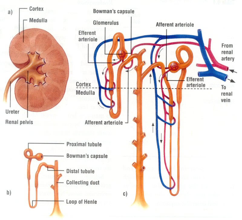
The collecting duct spans both the cortex and the medulla. In the bottom section (in the medulla), because the outside is hypertonic, even more water can be extracted (follows rules of osmosis). Hormones tell the collecting ducts how porous to make their membranes. If a membrane is made very porous, more water is absorbed into the medulla and the urine becomes even more concentrated. If the membrane is made not-very-porous, less or no water is absorbed so the urine is less concentrated.

2.70 Describe the structure of the urinary system, including the kidneys, ureters, bladder and urethra


Source: CrashCourse, YoutubeFigure 1

The urinary system is made up of:

  • the kidneys
  • the bladder
  • the ureter
  • the urethra
Looking at figure 1, you can see where each organ / part is located in the body. The functions of each part are below:
  • Kidney: filters out blood to prevent dehydration and manage the soluble substances in your blood and turns unwanted substances into urine
  • Bladder: stores urine
  • Ureter: carries urine from kidneys to bladder
  • Urethra: carries urine from the bladder to outside the body
Click here to see a video on the urinary system (seriously, it's worth it)

Friday, April 1, 2016

2.69 Understand how the kidney carries out its roles of excretion and osmoregulation

Nephrons in the kidneys (contains millions of nephrons) are in charge of excretion and osmoregulation.

Excretion
Excretion is filtering the bad stuff from the body and keeping the good bits we need. The kidneys excrete water, salts and urea from the body.

The liver converts amino acids (which contain nitrogen, which is toxic to the body) into urea. The kidneys filter urea from the blood stream and combine it with water to create urine which then moves into the bladder via the urethra.

Osmoregulation
Osmoregulation is the balancing of water levels in the body (remember OSMO-sis? Therefore water-regulation)


The kidneys react to ADH hormone released by the pituitary gland. 


Basically, in the nephron, there is a collecting duct. If more ADH is released, the walls of the duct will become more permeable and more water will be re-absorbed. If less, duct will become more impermeable.

2.68 Recall that the lungs, kidneys and skin are organs of excretion

Lungs
What? The lungs excrete carbon dioxide, which is a waste product of respiration. It diffuses into the lungs and is breathed out. 

Why? Carbon dioxide diffuses into blood plasma and makes it more acidic. It needs to be excreted so it doesn't reach toxic levels.As blood flows through the lungs, the carbon dioxide diffuses from the red blood cell into the alveoli and is breathed out.


Higher tier

If the levels of carbon dioxide are too high, the brain will detect this. It sends signals to increase the breathing rate so carbon dioxide can be removed from the blood more quickly, and the levels return back to normal.


Figure 1


Kidneys

What? The kidneys excrete excess water, salt and urea through urine. 

Why? (urea) Proteins are broken down into amino acids. Excess amino acids are broken down to form ammonia in the liver. It's then converted into urea (still in the liver). Although it is less toxic than ammonia, it still needs to be excreted from the body. 


(water) Water is a waste product of respiration and is also taken in by food or drink. Excess water could cause the plasma to be too dilute and cause blood cells to swell and burst :( while too little water could cause the plasma to be too concentrated and the blood cells to shrivel up / shrink and stop working. It's important to keep the water concentration in the blood plasma constant.


Skin

What? The skin excretes urea, water, salt and ammonia. This is released through sweat, which is a combination of urea, salts and water and is produced by the sweat glands.

Why? Things like ammonia and urea could be toxic if left in the body for too long. (see why water needs to be excreted by the kidneys)


Structure of the kidney: see 'pages' menu on right hand side

Source: BBC Bitesize http://www.bbc.co.uk/schools/gcsebitesize/science/triple_ocr_gateway/the_living_body/waste_disposal/revision/4/


Monday, January 25, 2016

2.62 Understand that platelets are involved in blood clotting, which prevents blood loss and the entry of micro-organisms

BACKGROUND INFO
- when you cut yourself / are injured, you are at risk of losing blood
- platelets are fragments of cells
- platelets are produced in the bone marrow

EVERYTHING IS EXPLAINED :D
Platelets release chemicals when there is an open wound that turn fibrinogen into fibrin, which is a solid. This forms a mesh which traps red blood cells. It eventually dries over to form a scab, and underneath, the tissue begins to repair itself.

(life hack - if you want a wound to heal quickly, don't try to pull of the scab!!)
Figure 1: A lovely picture of trapped red blood cells under the mesh
of fibrin - view from a microscope :)


IN SUPER SIMPLE STEPS
P - Platelets release chemicals
FiFi - Fibrinogen turns to fibrin
M - Mesh is formed
T - Traps red blood cells
D - Dries over
S - Scab forms
R - Repair happens in tissue underneath scab

HOW TO REMEMBER THAT
Papa Fifi makes tiny dark snakes run

(whatever works for you, really)

OR 
Here's an embarrassing song which might actually help:
*to the tune of twinkle twinkle little star*

Platelets release chemicals
Fibrinogen turns to fibrin.
Mesh is formed,
Traps red blood cells
It dries over
Scabs are formed
Tissue begins to repair
*insert final line here*

Again, whatever works for you. I was just...having a musical moment. Great. Now I've put it out there for the internet to see. Oh well, if it works, it's worth it :)


2.61 Understand that vaccination results in the manufacture of memory cells, which enable future antibody production to the pathogen to occur sooner, faster and in greater quantity

Vaccination in 5 simple steps!


  1. Person is injected with a weak or harmless sample of the pathogen / disease^
  2. Body fights off the disease as usual**
  3. Lymphocytes split, forming memory cells
  4. Upon re-infection*, the body produces antibodies much quicker, sooner and in greater quantity^
  5. Body develops 'immunity'^
(the ones that have a ^ beside it, see below for exam tips.)

Exam tips :) :)
 1. I reccommend using the word "pathogen" 
2. THESE ARE KEY WORDS - VERY VERY IMPORTANT TO MENTION! (the bold bit in step 4)
3. I'm not sure it's a great idea to say your body is immune because sometimes the disease does come back, or something very similar.

Extra words just for show ;)
** (step 2) this is the primary immune response.
* (step 4) this is the secondary immune response.

Figure 1 : Tee hee, hope you're okay with needles! :P


2.60 Describe how the immune system responds to disease using white blood cells, illustrated by phagocytes ingesting pathogens and lymphocytes releasing antibodies specific to the pathogen

What are white blood cells?
They are specialized cells that protect your body from pathogens (which can be harmful and cause disease)

I accidentally explained this in a previous post but I'll do so again:

Phagocytes
These types of white blood cells detect pathogens through a chemical trail that they leave behind. Their cytoplasm forms feelers called pseudopodia  which engulf the pathogen and forms a vacuole. The phagocyte releases digestive enzymes into the vacuole which then kill / digest it (the pathogen)

This process is called phagocytosis.
Figure 1
Lymphocytes
These white blood cells stand back a bit compared to the phagocytes (don't say that in an exam, for the love of elves, don't!). They release antibodies which are specific / complimentary to the antigens on the surface of the pathogen (also how it recognizes it). There are various types of lymphocytes - some cause the pathogens to stick together so phagocytes can engulf them all (the string of pathogens, I mean) , others disable / neutralize the cell and others latch onto the pathogen to attract phagocytes. When a lymphocyte encounters a pathogen and produces an antibody specific to that pathogen, it splits - one of the cells becomes a memory cell (See how vaccination works) and the other releases antibodies.


(because for some reason when I tried to put an image in, it didn't work)


Basic points on memory cells

  • Memory cells created when lymphocyte splits
  • Allows immune system to "remember" the disease (do not say that in an exam!!)
  • Upon re-infection, immune response is faster (secondary immune response is faster)
  • When the organism is re-infected, antibodies are produced much much quicker
:)

Lymphocyte types summary
  • Some cause pathogens to stick together (so phagocytes engulf them all)
  • Some latch onto them to attract phagocytes
  • Some neutralize the cell (or disable it, same thing)
Some also make pathogens explode but I'm not sure about that one so better not mention that in an assessment, be it an exam or just a test. Basically, do not mention that in an exam unless you have researched further and know how it works.

Little extra from Annabel: Just write all four things in an exam, even if it is a 3 mark question because you never know with the mark scheme... :)


2.66 Understand the general structure of the circulation system to include the blood vessels to and from the heart, the liver, the lungs and the kidneys

Quick overview of the meanings of some words:
Lung- pulmonary
Liver- hepatic
Kidney- renal
Stomach- gastric
Between the gut and liver is the hepatic portal vein.


(sorry if the diagram is a bit messy, it's hand made but hopefully accurate)