Monday, October 26, 2015

2.37 Describe experiments to investigate the evolution of carbon dioxide and heat from respiring seeds or other suitable living organisms

Experiment would be set up as follows to investigate release of carbon dioxide:

  • Put x grams of germinating seeds in a test tube.
  • Cover it with a bung/cork with a tube through it
  • Have one end of the tube in limewater
  • If it turns cloudy, carbon dioxide has been given off
  • See figure 1
Figure 1: Apparatus
To investigate the release of heat:

  • Put seeds in a test tube
  • Seal it with a cotton puff
  • Place a thermometer in it
  • Place the setup in a cold room (no less than 15ÂșC)
  • Measure initial temperature
  • Measure temperature after x amount of time (eg 20 mins)
  • Have another test tube, in the same room, with the same amount but have the seeds boiled. Leave it for the same amount of time.
  • Compare test tube 1 and 2. :)

Figure 2: Apparatus
Note that all images used in this post were created by me.

2.48 Describe experiments to investigate the effect of exercise on breathing in humans.

When you exercise, sometimes you end up respiring anaerobically as the body cannot get oxygen around quickly enough. Of course, this depends on the intensity of the exercise. This could be investigated as follows:

  • Ask a friend to volunteer 
  • At rest, have them lie down on the ground (belly up) and count how many times their chest rises in one minute. Record the result.
  • Now, ask them to walk 100m (or any other given distance). Measure their breathing rate the same way as before. 
  • Then ask them to run 100m. Measure breathing rate again.
  • Finally, ask them to sprint 100m. Measure their breathing rate again.
  • Wait until the person's breathing rate has returned to normal. Repeat the experiment from the start 3 times.
  • Take an average of the results
  • Plot a graph of your results
  • Remember to keep the person, distance run, and environment the same.
Dependent variable: Breathing rate (breaths per minute)
Independent variable: Intensity of exercise
Control variables: Distance (100m), person doing the exercise (fitness must be the same), environment (i.e not have one in super hot conditions and another in freezing cold)

Note: results that differ by more that 0.2 should be ignored and should thus not be taken into consideration when taking an average.

An example graph can be seen below. THIS GRAPH DOES NOT EXPRESS THE RESULTS OF THE EXACT EXPERIMENT DESCRIBED but it does express the same idea.
Conclusion: As exercise intensity increases, the breathing rate increases as well.

Sometimes, the image doesn't show up, so the link to it can be found here


2.46 Explain how alveoli are adapted for gas exchange by diffusion between air in the lungs and blood in capillaries


The alveoli are adapted to the job of making gas exchange quick and effective because (they have...) :
  • Walls one cell thick make diffusion quick and easy
  • There are a lot and they look a bit like bunches of grapes, meaning an enormous surface area - this means the blood stream is in contact with a large surface, and gas exchange happens quickly
  • Moist lining helps gases dissolve and diffuse into it
  • Constant blood flow allows all the blood to be oxygenated effectively
Figure 1: Diagram :)

2.45 Understand the role of the intercostal muscles and the diaphragm in ventilation

Intercostal Muscles
As you breathe in...

  • The muscles contract
  • The ribs move up and outward
As you breathe out...
  • The muscles relax
  • Ribs move in and down
Diaphragm
As you breathe in...
  • The diaphragm contracts, moving down
  • Low pressure is created inside the lungs,  forcing air in
  • Diaphragm pulls down
As you breathe out...
  • The diaphragm relaxes 
  • Pushes upwards
  • High pressure is created inside the lungs, forcing air out
Source: BBC BitesizeFigure 1: A diagram of the respiratory system


2.44 Describe the structure of the thorax, including the ribs, intercostal muscles, diaphragm, trachea, bronchi, bronchioles, alveoli and pleural membranes

How air travels...
Mouth/nose→Trachea→Bronchi→Bronchioles→Alveoli→Gas exchange occurs and then the process happens in reverse (i.e. from alveoli to bronchioles)

The functions of each of these parts:

Ribs - These are curved bones that embrace and protect the lungs from damage. They are linked together by intercostal muscles.

Intercostal muscles - Located between the ribs and expand and contract as the lungs fill or deflate. They keep the ribs in place too.

Trachea - Lined with C shaped (or U shaped, depends how you think of it) rings of cartilage, these protect it from being crushed. This is the pipe that air travels down from the mouth or nose.

Bronchi - The two tube like structures that divide the trachea, one leading into each lung. 

Bronchioles - Smaller tube like structures that contain alveoli at the tips and lead the air in and out of them

Alveoli - Tiny, grape-like air sacs surrounded by capillaries where gaseous exchange occurs

Pleural membranes - The outer lining of the lungs, which would feel kind of wet, and stops the lungs sticking to the ribs, as well as reducing friction. 

Figure 1: A diagram of the human thorax

2.40 Understand that respiration continues during the day and night, but that the net exchange of carbon dioxide and oxygen depends on the intensity of light

Figure 1: Plant during the day
Yes, plants photosynthesize, but they respire too. They respire throughout the day and the night, because, of course, it is a living organism. As you should know, glucose is used in respiration, which the plant produces through photosynthesis :)

During the night, a plant only respires, as there is no sunlight. This means that it gives off carbon dioxide and does not re-absorb it. Similarly, during the night, oxygen will not be given off, as the plant is not photosynthesizing.

Remember: photosynthesis depends on light, but respiration never stops ^_^

Figure 2: Plant at night


2.39 Understand gas exchange (of carbon dioxide and oxygen) in relation to respiration and photosynthesis

In both photosynthesis and respiration, gas exchange occurs: one gas is swapped for another. They are the opposite of each other, which is why it works.

photosynthesis ~ carbon dioxide + water → glucose + oxygen
The plant, or whatever is photosynthesizing, is using the waste products of respiration, taking in carbon dioxide and giving out oxygen as a waste product.

respiration ~ glucose + oxygen → carbon dioxide + water
The organism is using the products of photosynthesis (although glucose can be obtained through food, so I guess that doesn't really work..... you probably shouldn't put that in your exam...sozzies), taking in oxygen and giving out carbon dioxide.

Because why not, here is a diagram

2.38 Understand the role of diffusion in gas exchange

Diffusion is the random movement of particles from an area of high concentration to an area of low concentration. This is exactly what happens in gas exchange. 

It occurs in the alveoli, in the lungs, where oxygen is transferred into the bloodstream and carbon dioxide diffuses out of the blood and is exhaled. 


There is a high concentration of oxygen in the alveoli and a low concentration in the deoxygenated blood coming into the lungs. That is how diffusion occurs.

There is a high concentration of carbon dioxide in the blood coming into the lungs, which diffuses back into the alveolus and gets exhaled out of the body. 
↓↓↓
The gases move across the wall of the alveolus. 

Figure 1: A pulmonary alveolus


2.36 Write the word equation for anaerobic respiration in plants and in animals

In animals, it is:

glucose → lactic acid (+ energy)

In plants, it is:

glucose → ethanol + carbon dioxide (+ energy)

2.35 Write the word equation and the balanced chemical symbol equation for aerobic respiration in living organisms

Glucose + oxygen → carbon dioxide + water (+ energy)

C6H12O6 + 6O2    →    6CO2 + 6H2O (+ energy)

2.34 Describe the differences between aerobic and anaerobic respiration

Aerobic respiration is when an organism respires using oxygen and glucose is completely and fully broken down. This is used for movement, growth, etc. in humans. It is how we respire most of the time. This may be different in other organisms, or the same.

glucose + oxygen → carbon dioxide + water (+ energy)

Anerobic respiration is when an organism respires without using oxygen. Yeast, for example, naturally respires anerobically. In humans, however, we respire anerobically when the heart and lungs cannot work fast enough to provide enough oxygen around the body to break down that glucose. Instead, the body does not use oxygen, providing a fast, short burst of energy. This causes the build up of lactic acid (a waste product) in our muscles, which sometimes makes them sore. After respiring anerobically, as a human, you breathe heavily to make up for the oxygen lost. This can occur in other organisms too but may be different.

glucose → lactic acid (+ energy)


Figure 1: When you sprint, you respire anerobically. (Yes, that is Usain Bolt.)

2.33 Understand that the process of respiration releases energy in living organisms

All living things respire. (not to be confused with breathing) This occurs in the mitochondria of every cell, and produces energy for the organism. Even plants do this. It is the breaking down of glucose to produce energy.


2.47 Understand the biological consequences of smoking in relation to the lungs and the circulatory system, including coronary heart disease


  • Smoking causes tar to build up in your lungs, which can lead to cancerous mutations and the formation of tumors
  • It destroys the cillia that line the trachea (whose job is to stop unwanted particles entering the lungs)
  • It causes the build up of mucus (can cause smoker's cough) which could lead to infections in the trachea and bronchi; bronchitis
  • Hardens the arteries, which makes blood flow increasingly difficult and puts strain on the heart, leading to coronary heart disease.
Figure 1

2.17 Describe the process of photosynthesis and understand its importance in the conversion of light energy to chemical energy

Photosynthesis is the process by which light energy is converted into chemical energy, typically by a plant. Carbon dioxide and water is made into glucose. Oxygen is a waste product of the reaction.

carbon dioxide + water → glucose + oxygen

Photosynthesis is the reverse of respiration, so plants use the waste products of other animals to create "food" for themselves. This allows them to live. Plants are at the bottom of the food chain, as they don't "eat" like animals do; they harness energy from the sun. Because of this, other animals eat them, which bigger animals eat, etc - it forms a food chain.


Here's a picture of a plant to make your day :)

2.16 Describe experiments to investigate diffusion and osmosis using living and non-living systems

Diffusion

  • Fill a gas jar with water
  • Put a small quantity of strongly colored, solube solid at the bottom (you could use a straw for this)
  • Time how long it takes for the liquid to reach equilibrium (until it is all the same color)
  • You might want to repeat this at different temperatures of water, but that would be a separate investigation. However, science says that the higher the temperature, the faster diffusion will occur as the particles have more energy and move around faster :)
Osmosis
  • Cut two cubes of potato, about 2x2x2 cm or something similar
  • Weigh each one. Record their initial mass
  • Place one in a concentrated sugar solution (note that salt water can also be used) and the other in distilled water (see figure 1), making sure that they are placed in the same amount of solution.
  • After a set amount of time (let's say, an hour, for example), take them out and pat them dry with a tissue
  • Weigh each one again. Record their new mass
  • You should see that the potato's mass (in the sugar solution) would have decreased - this is because water moved through osmosis OUT of the potato. The potato's mass (in distilled water) would've increased, as water moved INTO the potato.
Figure 1

2.11 Describe experiments to investigate how enzyme activity can be affected by changes in temperature.

As you should know, the test for starch is to put iodine on a sample, and it will turn blue black if starch is present (or it may turn another color but the exam answer is blue black). To investigate how temperature affects the enzyme rate of reaction, you could do the following experiment:


  • Put x amount of starch in a test tube (eg 10 cm3) 
  • Add amylase to it
  • Put the mixture into a waterbath at temperature y
  • On tiles with little dimples (WHAT IS THAT CALLED?!), use a dropper/pipette to put a small sample of the mix (of what is in the test tube) onto one of the 'dimples' and add iodine to it. Do this every thirty seconds.
  • Time how long it takes for the samples to stop turning blue black
  • Repeat everything again, but at at different temperature. An example would be do do the experiment at 15ÂșC, 25ÂșC, 35ÂșC, 45ÂșC and 55ÂșC (in other words, you change temperature y)
  • Plot a graph
Figure 1: What your graph would look like (something similar)

2.10 understand how the functioning of enzymes can be affected by changes in active site caused by changes in pH

If the pH is too far from the ideal, optimum pH, the enzymes can de-nature, as a change in pH can cause the bonds that hold the structure in place to break :( This would mean that the substrate no longer fits in the active site. 

Different enzymes will work at different pH values. Eg the enzymes in your stomach will work best at a more acidic pH than those in your small intestine.

Figure 1: A graph showing the relationship between rate of enzyme activity
and pH

2.32 Describe an experiment to investigate the energy content in a food sample

Figure 1: Apparatus (but is missing the thermometer in the boiling tube)
  • Get 5 different food samples. For this experiment, let’s say crackers, apple, cornflakes, dried tomato and tofu. (We don't want to be killing animals...no thanks!)
  • Prepare the following apparatus: A utility clamp holding a boiling tube with a thermometer in it. Have a skewer and a bunsen burner at the side.
  • With each food sample, put it on the skewer and take the SAME amount and set fire to it. Place it directly underneath the boiling tube filled with water. Record how much the temperature has risen.
  • Empty the tube and take the SAME amount of water as before at the SAME (room) temperature. 
  • Repeat steps 3 and 4 for all the food samples. Now calculate the difference in temperature.
  • The more the temperature rose, the more energy the food sample has. :) To plot a graph, plot a bar graph with the x axis labelled with the types of food you used and the y axis labelled with percentage % change in temperature (yay…more calculation…)


2.31 Describe the structure of a villus and explain how this helps absorption of the products of digestion in the small intestine


These are tiny little hair-like things that line the wall of the small intestine and are responsible for the absorption of nutrients. (they are not actually hairs, just to make it clear)

  • Lined with microvilli → increased surface area → quicker diffusion
  • Blood capillaries → concentration gradient constant → nutrients (glucose & amino acids) quickly absorbed into bloodstream 
  • Walls one cell thick → quick diffusion → molecules can pass easily
  • Lacteal → central vessel in villi → responsible for absorption of fats


Figure 1: Structure of a villus


2.30 understand that bile is produced by the liver and stored in the gall bladder, and understand the role of bile in neutralising stomach acid and emulsifying lipids

Basically...
  • Bile is produced by the liver
  • It is used to neutralise the food after it has left the stomach, as it is highly acidic.
  • It emulsifies lipids, as they are not soluble in water: instead of one big oily blob, it is broken down into various tiny little blobs.
  • It is a gross shade of vomit green (but you don't need to know that)
Figure 1
(If you're a bit squeamish like I am, may I suggest that you do not look up "bile" on google images)


2.29 Understand the role of digestive enzymes, to include the digestion of starch to glucose by amylase and maltase, the digestion of proteins to amino acids by proteases and the digestion of lipids to fatty acids and glycerol by lipases.

Enzymes, as mentioned before, are biological catalysts. They help break down the food that moves through our digestive system. Different enzymes do different tasks, and break down very specific components of food:

amylase and maltase break down starch into glucose

protease breaks down proteins into amino acids

lipase breaks down lipids into fatty acids and glycerol 

Figure 1: The structure of lipids

2.28 Explain how and why food is moved through the gut by peristalsis

Food moves down the oesophagus through a process known as peristalsis. This is when the two sets of muscles in the gut contract, creating a squeezing action that moves the food down. 

This is necessary because food needs to be mechanically moved to get through the digestive system. 

Source: BBC BitesizeFigure 1: Peristalsis 

2.27 Understand the processes of ingestion, digestion, absorption, assimilation and egestion

Ingestion: the act of taking food into the body; eating

Digestion: the process by which food is broken down and nutrients are absorbed into the bloodstream. Large, insoluble molecules are broken down into smaller, soluble ones.

Absorption: the process by which the soluble molecules are taken from the gut (typically in the small intestine) and transported into the bloodstream for use in the body.

Assimilation: the act of absorbing molecules through the cells in tissues


Egestion: To get rid of as a waste product (undigested food. Not to be confused with excretion)

Figure 1: Part of the digestive system

2.26 Describe the structures of the human alimentary canal and describe the functions of the mouth, oesophagus, stomach, small intestine, large intestine and pancreas

I hope you like reading :)

Food goes from… the mouth → oesophagus (gullet) → stomach → small intestine → large intestine → anything that is not digested goes out the body through the anus

The mouth mechanically breaks up the food into physically smaller bits. This is then moved down the oesophagus through a movement known as peristalsis. From there, it goes through the stomach. The sphincter between the end of the oesophagus and the stomach opens as the food goes through. The (very strong) acid in the stomach further breaks down the food chemically. From there, it goes into the duodenum where pancreatic juices (from the pancreas) are added to the food and digestive enzymes are also secreted by the pancreas, which speed up the breaking down of the food. Bile is added to neutralise the “mixture”, as it was highly acidic after leaving the stomach. The nutrients from the food are absorbed in the small intestine thanks to the hundreds of thousands of villi that line the small intestine walls, whose large surface area helps the nutrients dissolve quickly into the blood stream. From here, any remaining food goes through the large intestine, where the water is absorbed. Any undigested substances are excreted.

Figure 1: Human digestive system

2.25 Understand that energy requirements vary with activity levels, age and pregnancy.


A bodybuilder is going to need a lot more energy (from proteins and carbs, and lipids) than a female 14 year old. A baby is going to need quite a bit of energy, as he/she is growing, but nowhere near as much as an adult. A pregnant woman is going to need a lot of energy, as she needs to supply enough chemical energy for herself and her baby.

Figure 5-1: Read the title. Also, sorry it's hard to read.

2.24 Identify sources and describe functions of carbohydrate, protein, lipid (fats and oils), vitamins A, C and D, and the mineral ions calcium and iron, water and dietary fibre as components of the diet

Carbohydrates 
Sources of simple carbs: Candy, fruit, etc.
Sources of complex carbs: Bread, pasta, beans, etc.

Carbs are the body’s main source of energy. Used as immediate energy.

Proteins
Sources: Beans, lentils, soy, etc. (I’m a vegetarian so I prefer to list that kind of stuff, but if not, protein can also be found in animal meat, including fish)

Protein is used to build and repair tissue and build muscle.

Lipids
Sources: oil (eg olive oil), fish, eggs, pork

Lipids are usually stored as fat for insulation and used as a long term energy store, as well as to protect (especially vital) organs.

Vitamin A
Sources: Green and yellow veggies (eg lettuce) and fish liver oil (yucks!) and dairy such as milk or eggs

Used to maintain eyes healthy, normal reproduction, formation and maintenance of teeth, skin and tissue. It is an antioxidant so it also helps the immune system function correctly.

Vitamin C
Sources: Oranges, lemons, raspberries, broccoli etc.

This is an antioxidant in the body and is essential in maintaining healthy connective tissue (deficiency causes scurvy) Also helps the absorption of iron and copper and helps fight and prevent infection.

Vitamin D
Sources: Sunlight, fish and liver oils. Few foods contain significant amounts.

Helps the body absorb calcium. (deficiency causes rickets)

Calcium
Sources: Milk, cheese, yoghurt and fortified soy milk, etc

Essential for maintaining strong bones, teeth and nails, functioning heart, muscles and nerves.
Iron
Sources: whole meat cereal, chickpeas, lentils, sunflower seeds, kale.

Used in haemoglobin in red blood cells, responsible for transporting oxygen around your body

Water
Sources: WATER!! (and soup lol)

Used in respiration.

Dietary fiber
Sources: Cereals, beans (eg chickpeas, red kidney beans, etc) and nuts


Important for healthy bowel function and reduces the risk of developing bowel cancer.

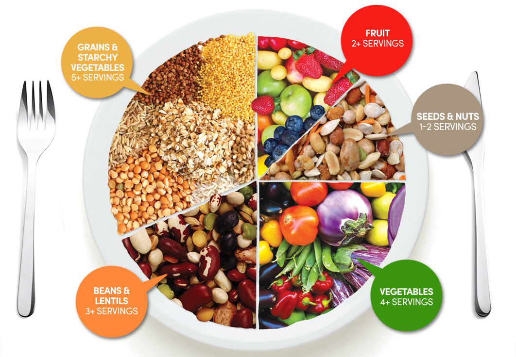
Figure 1: Vegetarian food plate, because why not
(yes, I am vegetarian...#sorrynotsorry)

2.23 Understand that a balanced diet should include appropriate proportions of carbohydrate, protein, lipid, vitamins, minerals, water and dietary fibre

A balanced diet should include correct proportions of carbs, proteins, lipids, vitamins, minerals and dietary fiber. This can be represented visually in the food pyramid. 

Figure 1: Food pyramid


For example, if a diet is too high in lipids and simple carbs (sugars), it is likely that the person will gain weight in an unhealthy way and experience health problems, such as clogged arteries due to excess cholesterol.

2.20 Describe the structure of the leaf and explain how it is adapted for photosynthesis

Figure 1: Labelled leaf diagram

  • Large surface area means more sunlight is absorbed
  • Usually very thin, meaning diffusion of carbon dioxide into the leaf can happen quickly
  • The stomata open and close, letting carbon dioxide in.
  • The epidermis is very thin and completely transparent, so all the sunlight can get to the leaf
  • There is a waxy cuticle that prevents water from getting into the leaf and reduces water loss
  • The waxy cuticle is also transparent to allow sunlight through
  • Palisade mesophyll is packed with chlorophyll and close to the top of the leaf so it gets the most sunlight, making photosynthesis more effective
  • In the spongy mesophyll there is quite a lot of air space which allows carbon dioxide to diffuse into the cells.

2.19 Understand how varying carbon dioxide concentration, light intensity and temperature affect the rate of photosynthesis


Concentration of CO2 (carbon dioxide)
If there is a higher concentration of CO2, the plant will photosynthesise more until a point where the amount of CO2 outweighs the rest that is needed for this. For example, if you put a plant in a room with a high concentration of CO2, it would photosynthesise more, but if you continued to increase the concentration, it would need more sunlight, or water, etc. to be able to continue increasing the rate at which is photosynthesizes. If the concentration is too low, the rate will decrease.

Light intensity
If the light / sunlight is more intense, the rate of photosynthesis will also increase. However, if the light is too bright, it might burn the plant’s leaves or cause it to overheat, in which case its enzymes would denature and the rate would go down :( . If the light is too dim, the rate of photosynthesis will decrease as it will not have enough light energy to transfer into chemical energy.

Temperature

If the plant is at optimum temperature, its rate of photosynthesis will be the highest. This is because the enzymes, that are biological catalysts and therefore speed up the plant’s metabolic reactions, will be working at their best. If the temperature is too high, the plant could burn, or its enzymes will denature, bringing down the rate of photosynthesis. Until that point, the higher the temperature, the higher the rate, as it has more energy from the heat .

Figure 1

2.18 write the word equation and the balanced chemical symbol equation for photosynthesis

Word: carbon dioxide + water → glucose + oxygen

Symbol: CO2 + H2O → C6H12O6 + O2

Balanced: 6CO+ 6H2O → C6H12O+ 6O2

Figure 1: Perfection
Don't forget to include the light energy and chlorophyll on the arrow!

2.5 Identify the chemical elements present in carbohydrates, proteins and lipids (fats and oils)

Carbohydrates - made up of carbon, hydrogen, oxygen
Proteins - made up of nitrogen, carbon, hydrogen, oxygen, sulphurphosphorus 
Lipids - made up of carbon, hydrogen, oxygen

Figure 1: The 3 main elements present in all.

2.2 Describe cell structures, including the nucleus, cytoplasm, cell membrane, cell wall, chloroplast and vacuole


Cell structures
  • Nucleus - controls the cell's behavior
  • Cytoplasm - makes up most of the cell, a gelatine-like subtance
  • Cell membrane - a thin wall that keeps the cell together
  • Cell wall - a harder, outer wall that keeps the cell's shape and stops it from bursting when it is full of water
  • Chloroplast - allows the organism to photosynthesize: to produce glucose from water and carbon dioxide
  • Vacuole - Where cell sap is stored, along with other nutrients
Key
Yellowish orange = Features in both plant and animal cells
Green = Typically associated with plant cells
Pale purple = Typically associated with plant cells, but a small temporary vacuole can be found in animal cells too

Figure 1: An unnecessarily complicated comparison

2.3 Describe the functions of the nucleus, cytoplasm, cell membrane, cell wall, chloroplast and vacuole


(Also covered in 2.2)

Functions:
  • Nucleus - controls the cell's behavior
  • Cytoplasm - makes up most of the cell, a gelatine-like subtance. This is where the organelles are found.
  • Cell membrane - a thin wall that keeps the cell together
  • Cell wall - a harder, outer wall that keeps the cell's shape and stops it from bursting when it is full of water
  • Chloroplast - allows the organism to photosynthesize: to produce glucose from water and carbon dioxide
  • Vacuole - Where cell sap is stored, along with other nutrients
Figure 1 (yes I know, same image again. Sozzles.)


2.4 Compare the structures of plant and animal cells

Part of structure          Present in plant c.         Present in animal c.
Nucleus                                   Yes                                   Yes
Cytoplasm                               Yes                                   Yes
C. membrane                          Yes                                   Yes
Vacuole                                   Yes (permanent)              Yes (but not permanent)
C. wall                                     Yes                                    No
Chloroplast(s)                          Yes                                   No

Further comparison

  • In plant cells, the vacuole is large and permanent, while in animal cells, it tends to be small and is not permanent.
  • Animal cells will burst if they are filled with too much water (turgid) but plant cells will not (due to the cell wall)

2.6 Describe the structure of carbohydrates, proteins and lipids as large molecules made up from smaller basic units

(I cut out a bit of the spec. point again, sozzles. But I'm going to repeat it anyway.)

The structure of carbs, proteins and lipids are large molecules:

  • Proteins are made up of amino acids
  • Starch and glycogen are made up of simple sugar
  • Lipids are made of fatty acids and glycerol
Figure 1

2.7 Describe the tests for glucose and starch

Glucose
Put a few drops of benedict's solution in a test tube of the substance that you want to test for glucose and place it in a warm waterbath. Leave it for a few minutes. It will be blue, initially. If there is any change in colour, glucose is present.

Figure 1: Test for sugar scale
Starch
Put a drop of iodine solution on whatever you wan to test for starch (let's say, a cornflake). If there's a change in colour, starch is present.

Figure 2: The test for starch, using a potato as an example


2.8 Understand the role of enzymes as biological catalysts in metabolic reactions

Enzymes are biological catalysts for metabolic reactions. This means that they speed up the chemical reactions needed to maintain life. 

Eg digestive enzymes speed up the breaking down of food

Figure 1

2.9 Understand how the functioning of enzymes can be affected by changes in temperature, including changes due to change in active site

Enzymes work best at an optimum temperature. This can be different from organism to organism, but in humans, it is about 36 - 38 ÂșC

If the temperature is too low, the enzymes won't have enough energy to speed up the reaction(s) much.

If the temperature is too high, the enzymes begin to denature, meaning that their protein structures change. The shape of the active site changes and they can no longer collide with the right substrate, as it doesn't fit in the active site.

Figure 1

2.12 Understand definitions of diffusion, osmosis and active transport

Diffusion: The random movement of particles from an area of high concentration to an area of low concentration.

Osmosis: The random movement of water particles from an area of high water concentration to an area of low water concentration through a semi-permeable membrane

Active transport: The movement of ions or molecules across a membrane from an area of low concentration to an area of high concentration, assisted by enzymes and requiring energy.


2.13 understand that movement of substances into and out of cells can be by diffusion, osmosis and active transport

Movement of substances (water, ions, molecules, etc.) in and out of cells can happen through diffusion, osmosis, and/or active transport.

(point is pretty self explanatory)

2.14 Understand the importance in plants of turgid cells as a means of support

Plants need water to perform photosynthesis. Turgid cells are cells that are full of water. Due to the cell wall, it does not burst (unlike animal cells). The cytoplasm and cell membrane is pressed against the cell wall, making the cell look "inflated" (which, it technically is but it's not great to write that in an exam)

Figure 1


Because the cells are turgid, they press against each other, forming a firm structure, and so are a plant's means of support.


2.15 Understand the factors that affect the rate of movement of substances into and out of cells, to include the effects of surface area to volume ratio, temperature and concentration gradient

Temperature: The higher the temperate, the faster diffusion or osmosis occurs. If the temperature is too high, however (too far past optimum), enzymes begin to de-nature and this may affect the rate of substance movement, especially in terms of active transport. Until that point, the higher the temperature, the faster the movement of particles, as they have more energy, and therefore collide more, making it more likely that they will move into the desired space.

Concentration gradient: If the concentration gradient is high/steep, it means that there is a greater difference in concentration between the substance inside and outside the cell. A steeper concentration gradient means the substance will diffuse faster because molecules have a bigger opportunity of diffusing.

Surface area to volume ratio: If there is a larger surface area to volume ratio, the movement of the substance is likely to be quicker, especially in terms of diffusion, as there is a larger space that particles can get past.

Sunday, October 25, 2015

2.1 describe the levels of organisation within organisms: organelles, cells, tissues, organs and systems.

In order from smallest to largest....

Organelles

  • Have a specific function inside the cell
  • Made up of small molecules
  • Example: mitochondria, ribosomes, etc. 
Cells
  • The basis of all living things
  • Said to be a functional unit
  • Made up of organelles
  • Examples: red blood cells, white blood cells, etc.
Tissues
  • Made up of many cells
  • Cells serve a common function
  • Examples: muscle tissue, neural tissue
Organs
  • Made up of tissues 
  • Form a large functioning unit
  • Examples: liver, heart, etc.
Organ systems
  • Made up of many organs
  • Serve a common or related function in the body
  • Many organs working together
  • Examples: respiratory system, skeletal system, etc.
Figure 1