
Saturday, April 2, 2016
2.76 Understand that urine contains water, urea and salts.
Again, incredibly self explanatory. Urine is made up of mainly water, urea and salts that the body no longer needs. (also has organic and inorganic compounds)

2.75 Describe the role of ADH in regulating the water content of the blood
THIS FINALLY MAKES SENSE.
Excess or lack of water is detected in the brain by the hypothalamus. It causes the pituitary gland to produce ADH (anti-diuretic hormone). It travels through the blood stream and into the kidneys - specifically, into the collecting duct. According to the water levels in the body, it will give a signal to either make the membrane (walls) more porous. Increased levels of ADH are typically secreted when there is lack of water in the body, because it makes the membrane walls more porous.
The more porous the membrane is, the more water is absorbed.
It causes urine to be more concentrated and of less quantity. It is also typically a yellowish color. If there is too much water, the levels of ADH will lower and the opposite effects will happen; the membrane becomes less porous, less water is absorbed and urine is clear and less concentrated (and more in quantity)
FOR STUDYING
A ridiculously long and rather unhelpful acronym:
Brain detects water levels
Hypothalamus (does this)
Pituitary gland produces ADH
(B)lood stream carries hormone
(K)idneys receive hormone
(C)ollecting duct specifically
(M)membrane
Porous (refers to membrane)
Water
(A)bsorbed
Returned to blood stream
BHPBKCMPWAR (long version)
BHPPWR (simplified)
A very simplified acronym of the process:
Detect
Produce ADH
Carry
Recieve
Collecting duct
Porous (membrane)
Absorbed
Returned (to blood stream)
DPCRCPAR
(Daddy Paced Carol's Room Cindy Passed Away Restfully)
For the super long acronym, here is a suggestion for remembering it (refers to Harry Potter)
Brave
Harry
Painted
Blue
Kittens
Cursing
Merrily
Patently
Wearing
Red
Excess or lack of water is detected in the brain by the hypothalamus. It causes the pituitary gland to produce ADH (anti-diuretic hormone). It travels through the blood stream and into the kidneys - specifically, into the collecting duct. According to the water levels in the body, it will give a signal to either make the membrane (walls) more porous. Increased levels of ADH are typically secreted when there is lack of water in the body, because it makes the membrane walls more porous.
The more porous the membrane is, the more water is absorbed.
It causes urine to be more concentrated and of less quantity. It is also typically a yellowish color. If there is too much water, the levels of ADH will lower and the opposite effects will happen; the membrane becomes less porous, less water is absorbed and urine is clear and less concentrated (and more in quantity)
FOR STUDYING
A ridiculously long and rather unhelpful acronym:
Brain detects water levels
Hypothalamus (does this)
Pituitary gland produces ADH
(B)lood stream carries hormone
(K)idneys receive hormone
(C)ollecting duct specifically
(M)membrane
Porous (refers to membrane)
Water
(A)bsorbed
Returned to blood stream
BHPBKCMPWAR (long version)
BHPPWR (simplified)
A very simplified acronym of the process:
Detect
Produce ADH
Carry
Recieve
Collecting duct
Porous (membrane)
Absorbed
Returned (to blood stream)
DPCRCPAR
(Daddy Paced Carol's Room Cindy Passed Away Restfully)
For the super long acronym, here is a suggestion for remembering it (refers to Harry Potter)
Brave
Harry
Painted
Blue
Kittens
Cursing
Merrily
Patently
Wearing
Red
2.74 Understand that selective reabsorption of glucose occurs at the proximal convoluted tubule

Pretty self explanatory. Basically, glucose is re-absorbed in the proximal convoluted tubule (aka the first twisty tube). This is because glucose is needed in the body for respiration. It is removed in the nephron (which is the entire yellow structure shown in the diagram above) and taken back into the blood stream.
2.73 Understand that water is reabsorbed into the blood from the collecting duct
Water is re-absorbed in the collecting duct one last time into the medulla because it is so salty.
Following the rules of osmosis, water travels from a high concentration to a low concentration through a semi permeable membrane - that is exactly what happens, because the concentration of water in the medulla is low compared with the filtrate. However, it depends on how permeable the membrane(s) are/is: the more permeable, the more water is absorbed. I.e. if your body lacks water, the membrane will be permeable so there is no unnecessary water loss.
More about this on post 2.71
Following the rules of osmosis, water travels from a high concentration to a low concentration through a semi permeable membrane - that is exactly what happens, because the concentration of water in the medulla is low compared with the filtrate. However, it depends on how permeable the membrane(s) are/is: the more permeable, the more water is absorbed. I.e. if your body lacks water, the membrane will be permeable so there is no unnecessary water loss.
More about this on post 2.71
2.72 Describe ultrafiltration in the Bowman’s capsule and the composition of the glomerular filtrate
In your kidneys, there are thousands of little structures called nephrons. These are responsible for filtering the blood. It begins in the glomerulus.
The pressure in the glomerulus is high enough to squeeze some fluid out of the blood. This enters the Bowman's capsule and is known as the filtrate. It is made up of water, urea and smaller molecules and ions. But how exactly does this happen?
The blood comes in through the afferent arteriole at high pressure. It branches and becomes much smaller and the pressure increases further. The high pressure forces the plasma (liquid) into the space inside the bowman's capsule. It is now called filtrate.
 Glomerular filtrate is composed of mainly water, ions (potassium, calcium, chlorine, etc.) and organic molecules.

The pressure in the glomerulus is high enough to squeeze some fluid out of the blood. This enters the Bowman's capsule and is known as the filtrate. It is made up of water, urea and smaller molecules and ions. But how exactly does this happen?
The blood comes in through the afferent arteriole at high pressure. It branches and becomes much smaller and the pressure increases further. The high pressure forces the plasma (liquid) into the space inside the bowman's capsule. It is now called filtrate.


2.71 Describe the structure of a nephron, to include Bowman’s capsule and glomerulus, convoluted tubules, loop of Henlé and collecting duct
 |
| Figure 1 |
- the glomerulus
- the bowman's capsule
- convoluted tubules
- the loop of Henle
- the collecting duct
Blood enters the nephron through the glomerulus. Here, some liquid is 'squeezed' out of the blood due to the high pressure. This is known as the filtrate. Large particles / cells such as erythrocytes (red blood cells) and large amino acids are not filtered. Instead, they continue in small blood vessels. Glucose is also re-absorbed immediately into the capillaries. The filtrate goes into the bowman's capsule and that is where the filtering process begins.

The beginning is in the renal cortex and consists of the glomerulus and the bowman's capsule which surrounds it. The bowman's capsule is a cup-shaped structure that takes in the filtrate while the glomerulus is a 'knot' of capillaries in the middle. The bowman's capsule is the starting point for the filtrate.
The glomerulus is directly connected to the proximal tubule, which is the first convoluted tubule. It's basically a windy tube where mainly organic solutes are re-absorbed, along with water and things like glucose, amino acids, sodium and potassium among others.

After the Loop of Henlé, it reaches the distal tubule, which is the farther-away-curly-tube, aka the second convoluted tubule. Here, the levels of potassium, calcium and sodium are regulated. This is done by pumps and hormones. Once it is done, all of the wanted particles, water, salt, etc. have been taken out, which leaves excess water, urea and other types of metabolic waste.

The Loop of Henlé dips down into the renal medulla and is a hairpin shaped tubule. The upper section is in the outer medulla and the lower section in the inner medulla. It extracts mainly water as it travels down into the medulla. On its way up, it pumps out the salts that the body needs. This causes the medulla to become really really salty and creates a concentration gradient (medulla becomes hypertonic). The further down you go, the saltier it is. In the descending end, the membrane is highly permeable to water. Not really to salt or anything else. When the filtrate gets to the bottom of the loop (because the inner medulla is so so salty) it's highly concentrated. In the ascending end of the loop, the process happens in reverse. Here, the membrane is not-so-permeable to water and instead is lined with channels that transport ions like potassium, sodium and chlorine.
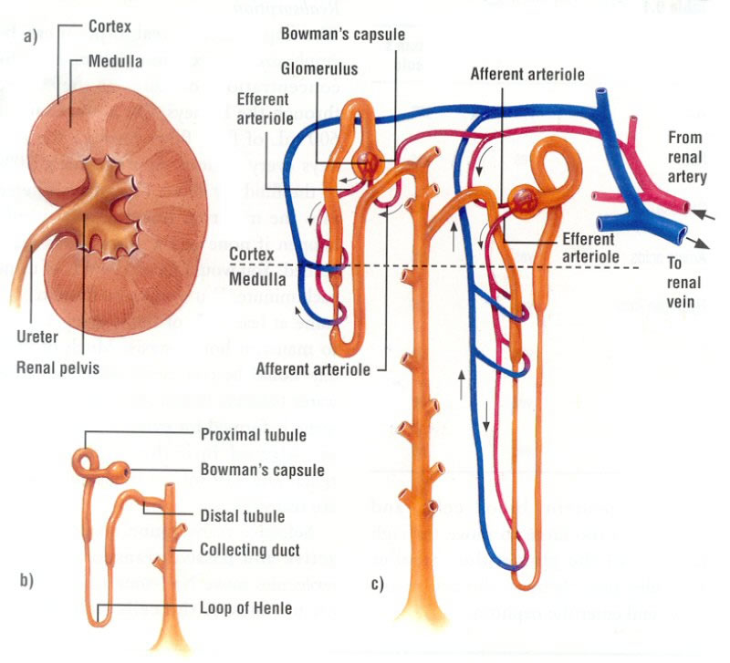
The collecting duct spans both the cortex and the medulla. In the bottom section (in the medulla), because the outside is hypertonic, even more water can be extracted (follows rules of osmosis). Hormones tell the collecting ducts how porous to make their membranes. If a membrane is made very porous, more water is absorbed into the medulla and the urine becomes even more concentrated. If the membrane is made not-very-porous, less or no water is absorbed so the urine is less concentrated.
2.70 Describe the structure of the urinary system, including the kidneys, ureters, bladder and urethra
 |
| Source: CrashCourse, YoutubeFigure 1 |

The urinary system is made up of:
- the kidneys
- the bladder
- the ureter
- the urethra
Looking at figure 1, you can see where each organ / part is located in the body. The functions of each part are below:
- Kidney: filters out blood to prevent dehydration and manage the soluble substances in your blood and turns unwanted substances into urine
- Bladder: stores urine
- Ureter: carries urine from kidneys to bladder
- Urethra: carries urine from the bladder to outside the body
Click here to see a video on the urinary system (seriously, it's worth it)
Subscribe to:
Posts (Atom)